Lateral side is convex medial is concave kidneys sit in a depression called the renal sinus. The _____are structures that separate the renal pyramids.
2
What is the medial indentation on the kidneys called.

. The kidneys lie in the retroperitoneal space of the posterior abdominal cavity. When viewing the internal anatomy of a kidney the outer region is known as the _____. The indentation on the medial surface of the kidney is called the _____.
A medial indentation where several structures enter or exit the kidney ureters blood vessels and nerves Adrenal Glands. The indentation is called the hilum. What is the outer tissue layer of the kidneys called.
Quiz 76 The prominent indentation on the medial surface of the kidney. The apex of a renal pyramid is called the. There is a deep indentation called the hilus on the medial side of each kidney from which the ureters and blood vessels enter and leave.
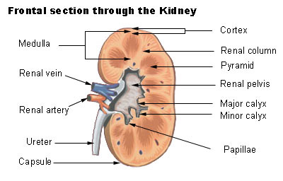
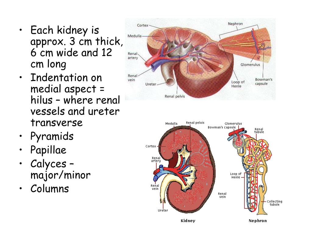
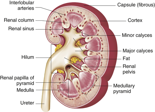
The medial margin of the kidney is concave region in the center called the renal hilum. It is roughly bean-shaped with an indentation called the hilum on the medial side. Emerging from the hilum is the renal pelvis which is formed from the major and minor calyxes in the kidney.
View Quiz 76docx from BIOL 1301 at Lone Star College System Woodlands. Each kidney has a medial indentation known as the renal hilum and an adrenal gland sits atop each kidney. Renal corpuscles and convoluted tubules.
This indentation is called the hilum and is the location where the renal artery and nerves enter the. Renal vein and ureter. Each kidney has a medial indentation known as the renal hilum and an adrenal gland sits atop each kidney.
The kidneys lateral border is convex whereas the medial border is concave and indented in a depression called the renal hilus. The renal hilum is the entry and exit site for structures servicing the kidneys. The outermost region is known as the _____.
The medial indentation where the ureter blood vessels and nerves are connected to the kidney is called the _____. The hilum leads to a large cavity called the renal sinus within the kidney. Renal veins artery nerves and lymphatic vessels are located in.
The kidneys are bean-shaped with the convex side of each organ located laterally and the concave side medial. Learn vocabulary terms and more with flashcards games and other study tools. The medial-facing hila are tucked into the convex indentation of the kidney.
List 5 structures that enterexit here. The indentation is called the hilum. The medial-facing hila are tucked into the sweeping convex outline of the cortex.
Entrance is called the HILUM-superior end of the kidney forms a funnel shaped sac - renal pelvis-renal medulla center of the kidney-renal cortex outer shell around the medulla. Urinary system made up of the kidneys ureters bladder and urethra. Each kidney has a medial indentation known as the renal hilum and an adrenal gland sits atop each kidney.
A renal capsule B renal column C renal pyramid D renal hilum. What glands sit on top of the kidneys. What is the medial indentation on the kidney called.
A gland that sits atop each kidney and is from the endocrine system. Figure 2512 Left Kidney. Where are the kidneys located in the body.
The medial indentation on each of the kidneys is known as the hilum also known as the hilus. Regions of the Kidneys x3 The regions where other structures exist and perform specific functions. What vessel branches off the aorta and brings blood into the.
The indentation on the concave side of the kidney known as the renal hilus provides a space for the renal artery renal vein and ureter to enter the kidney. Vessels nerves lymphatics and ureters. There are three regions of the kidney.
The lateral side of the kidney is curved outward with an indentation on the medial side. What is the renal cortex made of. All related structures enter or leave the kidney at the hilus.
Positioned behind the parietal peritoneum _____ lateral side is convex medial is concave indentation is the _____ entrance is called the _____. What tissue is found in the renal capsule. In the deep indentation on the concave side of the kidney the sinus.
What is the indentation on the medial side of the kidneys called and what emerges from this. A frontal section through the kidney reveals an outer region called the renal cortex and an inner region called the renal medulla Figure 2512. The cortex appears granulated due to the presence of nephrons.
Each kidney has a medial indentation known as the renal hilum and an adrenal gland sits atop each kidney. What is the posterior bladder. What is the function of the renal capsule.
What is inner tissue layer of the kidneys called. Lie on either side of the verebral column deep in the abdominal cavity. In the adult each kidney is approximately 3 cm thick 6 cm wide and 12 cm long.
What are the 3 regions that make up a kidney. What is the medial indention on the kidneys called.
Seer Training Kidneys

A P Lab Final Flashcards Quizlet

Renal Cortex Minor Calyx Renal Pyramid Arcuate Vein Major Calyx Ppt Video Online Download

Essentials Of Human Anatomy And Physiology 11e Marieb Chapter 15 The Urinary System Flashcards Quizlet
Ppt Urinary System Powerpoint Presentation Free Download Id 564837

Human Anatomy Physiology Ppt Download

Urinary System
Structure And Function Of The Renal And Urologic Systems Basicmedical Key
0 comments
Post a Comment